 Confocal Scanning Acoustic Microscopy (C-SAM)
Overview
Confocal Scanning Acoustic Microscopy (C-SAM) is a powerful imaging technique used in materials science, biology, and various industries. Unlike traditional optical microscopy, C-SAM uses high-frequency ultrasound waves to examine the internal structure, composition, and mechanical properties of materials at the microscopic level.
C-SAM works by focusing ultrasound waves on the sample, and by measuring the time it takes for reflected waves to return, it creates detailed cross-sectional images. The confocal aspect of C-SAM, achieved through a confocal aperture, enhances its ability to produce high-resolution images with excellent depth discrimination.
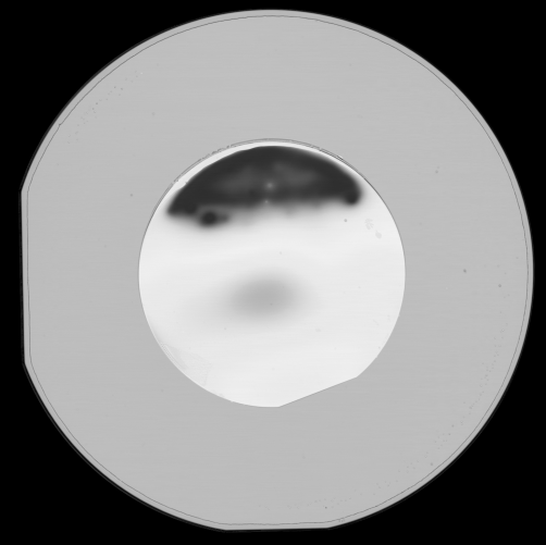
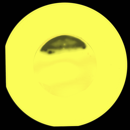
C-SAM provides critical information about subsurface defects, material properties, layer thickness, and even constructs 3D images. It has revolutionized fields like materials analysis, semiconductor inspection, and biomedical research, aiding in quality assessment and defect identification.
In summary, C-SAM combines ultrasound technology with confocal scanning to non-destructively examine the internal properties of materials, making it an invaluable tool for understanding complex structures in various disciplines.
Services
- Non-destructive analysis of delamination, voids, and cracking within structured samples
Pricing
- Regular service: Starts from $300 per sample with 4 – 6 days turnaround.
- Relaxed service: Starts from $200 with 7 – 10 days turnaround.
- For a comprehensive overview of our pricing structure, please log in to the Bee Portal.
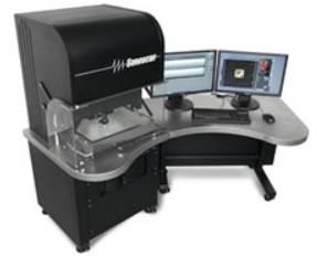
Equipment
- PolyGate™ technology with Multi-Gate™ and Probing-Gate™ functions capable of single and multi-focus imaging
- Up to 100 gates per channel with 2 Gsps sampling rate
- Windows® 7 Ultimate for multi-language and 64 bit capabilities
- Inertially Balanced Linear Motor Scanner with counterweight to minimize vibrations and ensure optimal scanning results
- Tower mounted scan reference platform and sample fixture
- Open-access scanning area makes loading and unloading easy and is capable of scanning JEDEC trays or a 300mm wafer
- Water recirculation and inline temperature control options available
- Virtual Rescanning Mode (VRM)™ stores comprehensive data and enables you to perform a complete analysis of a sample, even when it is no longer available
- SonoSimulator™ is integrated into the Gen6 to help image thin, multi-layer samples, such as Stacked Die
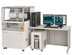
- Fine Tiny but still harmful flaws can no longer hide under FineSAT system where high performance ultrasound unit and precision scanner are perfectly designed in! Equipped with uncompromised 500MHz bandwidth electronics, high sensitivity transceiver and ultra-precise scanner mechanism, the resultant image definition can be as high as 8,192×8,192pixels at the finest pitch of 0.5µm.
- Fast Quick and easy acquisition with total data reproducibility! Max scanning speed 1,000mm/s, quick acquisition tools, and continuous auto-measurement features assure the highest productivity ever! Just one-click from ‘Image Index’ makes each and every acquisition perfect, and then move on to the characterization and judgment stages.
- Flexible Loaded with variety of analytical software tools! Waveform, intensity, depth, etc. acoustic wave analysis tools in the new software just get better! Data acquisition and image processing techniques with very high frequency probes and through transmission probes are greatly enhanced!
FAQ
A: The fundamental principle of C-SAM is the use of focused ultrasound waves. A specialized acoustic lens generates these waves, which are directed at the sample. The reflected waves are measured to create detailed cross-sectional images.
A: C-SAM is typically used in failure analysis cases in microelectronic parts. It is also used in biological and medical samples.
A: C-SAM is non-destructive technique as it uses sound waves to image the samples interfaces. The sample is typically submerged in water in which the sound travels through.
A: Resolution can go down to 1-3 um with a frequency of 400 Mhz.
A: Confocal Scanning Acoustic Microscopy (C-SAM) is an advanced imaging technique that uses high-frequency ultrasound waves to investigate the internal structure, composition, and mechanical properties of materials at the microscopic level.
A: C-SAM differs from optical microscopy in that it uses ultrasound waves instead of light to examine materials. This allows C-SAM to analyze both transparent and opaque materials and provides depth information within the specimen.
A: C-SAM offers non-destructive imaging, the ability to assess internal structures, and the detection of subsurface defects, making it invaluable for materials analysis, especially in composite materials and semiconductor manufacturing.
A: C-SAM can provide information about subsurface defects, material properties, the thickness of layers within a specimen, and even create 3D images by scanning through multiple focal planes.
A: C-SAM is used in various industries and fields, including materials science, semiconductor inspection, biology, and engineering. It aids in quality control, defect identification, and structural analysis.
A: In biomedical research, C-SAM can be used to characterize biological tissues, study cell structures, and assess biomaterials, providing insights into the mechanical properties and internal structure of biological samples.
A: C-SAM may have limitations related to sample size and preparation, and it may not provide information about certain materials, such as highly absorbing or attenuating samples. Additionally, it requires specialized equipment and expertise.
A: Yes, C-SAM can be used for quantitative analysis by measuring the time delays of reflected ultrasound waves and relating them to material properties or thickness. This allows for quantitative assessments of certain characteristics within a sample.